
3D printer
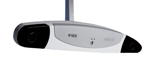
Polaris optical tracker

CNC machine

Laser cutter
Equipment
The following equipment is just a partial list of what is available at Sheikh Zayed institute ─ Children’s National for the execution of the proposed project.
Use this link to book equipment: https://app.clustermarket.com/login
Optical tracker
A number of optical and electromagnetic trackers and tracking accessories from Northern Digital, including a Polaris optical tracker (Northern Digital Inc., Waterloo) are available for conducting robot positioning accuracy study. Up to 15 wireless tools (maximum 6 active wireless) with a maximum of 32 passive and 32 active markers could be tracked with this optical tracking system. Volumetric accuracy of this system is 0.25 mm RMS.
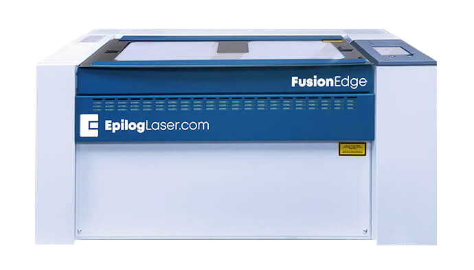
Laser cutter
A CO2 laser systems (Epilog Laser, CO, USA) is available for high-quality engraving and cutting of wood, acrylics, and plastics. This Mini Epilog Laser system has a workspace of 24`` x 12``. Its 40-watt laser beam is suitable for cutting acrylic and plastic sheets with 1/4`` thickness. The cutting depth could be increased to ½`` by adjusting laser focus.
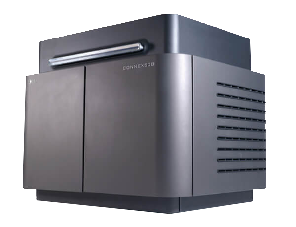
3D printer
A 3D printer/rapid prototyping machine (Objet Connex500) is available for building small mechanical parts/fixtures in-house. Using a variety of materials, this printer is able to build parts up to 19.3” × 15.4” × 7.9’’.
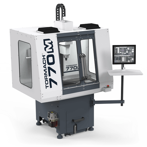
CNC mill
Tormach PCNC 770 is a 3-axis bench top CNC mill with a 1.0 HP spindle powered by a belt driven induction motor. The spindle speed is selectable through two ranges: 175 to 3,250 rpm in the low ratio, and 525 to 10,000 rpm in the high ratio. This broad selectable range of spindle speeds, offers the flexibility to machine materials ranging from soft plastics to ferrous metals. This machine will be used for building small mechanical parts as needed.
MRI Scanners
Interventional Cardiac Magnetic Resonance Suite: Constructed in 2013, the ICMR Suite at Children’s National houses a 1.5T Siemens Aera magnet co-located with a recently renovated Siemens cardiac catheterization laboratory with biplane fluoroscopy system and GE Maclab hemodynamic recording system. The magnet and fluoroscopy systems are equipped with two methods of image overlay software including Siemens X-ray Fused with MR (XFM) software, and Artis zee with PURE overlay software which allows for visualization of previously acquired CMR images during fluoroscopic image acquisition. The ICMR Suite is fully wired to receive input from the PRiME hemodynamic monitoring system which can be used in the MR scanner to monitor patients during their cardiac catheterization.
Philips Achieva 1.5T MRI scanner – This scanner is located in the operating rooms and available to the research team on an as-needed basis. There are several MRI technologists available to assist with experiments. The Children’s team has done many experiments with this system in the past.
CNH MRI suites house one 3T Discovery MR750 scanner (GE Healthcare, Milwaukee, WI) with an actively shielded superconducting whole body magnet with a 60cm bore with a detachable table, and equipped with a gradient system of 50mT/m amplitude and 200T/m/s slew rate, and 128-channel optical radio frequency receiver chain with 18 superconducting shim coils. The system software is version DV26x, with 2D and 3D whole body structural, functional, perfusion, diffusion imaging and multinuclear 1H, 31P, 13C, 23Na or 19F single- and multi-voxel spectroscopy acquisition methods. It is also equipped with post-processing application software for fMRI, ASL perfusion, diffusion and spectroscopy. Radio frequency coils available are a 32-channel receive-only head coil (MR Instruments, Inc., Minneapolis, MN), a 16-channel head-neck-spine receive-only coil array (USA Instruments Inc., Aurora, OH), an 8-channel Torso coil array, a quadrature transmit/receive brain coil, and general purpose receive-only flex coil arrays.
CNH Radiology also has two 1.5T Discovery MR450 scanners (GE Healthcare, Milwaukee, WI), one 60 cm another 70 cm bore (450W GEM), with actively shielded superconducting magnets, whole body, detachable table, MRI scanners, with a gradient system of 50 mT/m amplitude and 200 T/m/s slew rate, 32-channel optical RF-receiver chain with 18 superconducting shim coils. The system software is version DV26x, with 2D and 3D whole body structural, perfusion, diffusion imaging and 1H spectroscopy acquisition methods. Radio frequency coils available are an 8-channel receive-only head coil (Invivo, Gainesville, FL), a 16-channel head-neck-spine receive-only coil array (USA Instruments Inc., Aurora, OH), a 32-channel receive-only cardiac coil array (Invivo, Gainesville, FL), an 8-channel receive-only cardiac array, an 8-channel receive-only body array, a quadrature transmit/receive brain coil, and general purpose receive-only flex coil arrays.
GE MR450W GEM 1.5T MRI and Insightec ExAblate 4000 (clinical MRgFUS system) – ExAblate 4000 is a clinical MR-guided focused Ultrasound (MRgFUS) system for treatment of the brain, and is intended to be installed and configured to the GE MR450W 1.5T with GEM Suite. It is a transcranial MRgFUS system designed for the non-invasive ablation of brain tissue for the treatment of medication-refractory tremor in patients. ExAblate system treatment consists of accurately guide the focus of the ultrasound energy to the target region. Focused ultrasound energy is then repeatedly transmitted to the target until the desired outcome is achieved. Targeting is accomplished using magnetic resonance (MR) images taken during the treatment. The treatment process is constantly monitored by real-time closed-loop thermal feedback under full control of the treating physician. Once the targeting is complete, the treatment outcome is confirmed with MR imaging sequences immediately after the treatment.
ExAblate 4000 system is comprised of treatment table, operator’s console, equipment and water system cabinets, and front end electronics unit. The treatment table contains the focused ultrasound (FUS) transducer with its positioning system. It is a modified GE MRI table that docks to the MR system in the same way that the standard MR table docks, and has been adapted to be used with DV26 software version. During treatment, the patient lies on the patient table. The patient’s head is fixated to the table, using a standard stereotactic head frame. The table also contains the FUS transducer located on a mechanical positioning system, so that transducer can be moved to the desired location relative to the patient’s head. The table may be taken out of the magnet room either during patient preparation procedures or for storage when not in use. Front End (FE) electronic unit will be located in the magnet room and contains the electronic modules that drive the ultrasound transducer. The FE contains 1024 power amplifiers to drive the desired signal in each of the elements in the ExAblate 4000 system phased array transducer. The FE also contains control and monitoring electronics for the power amplifiers, as well as filters-amplifiers for acquisition of acoustic feedback for cavitation monitoring purposes. It is intended that the cabinet remains in the magnet room when not in use. ExAblate 4000 system includes a water system used for acoustic coupling between the patient and the ultrasound transducer. Clinical procedures and treatments require constantly circulating cooled water in interface between patient’s skull and the transducer. In-built body warming system is utilized to keep warm air flowing on the patient’s body to keeps body temperature and comfort at normal levels. ExAblate system is comprised of a cluster of 2 interconnected PC units – the FUS application workstation and the hardware control PC. In addition, FUS is connected to the GE MR system for online scan control and MR image retrieval.